MORE ABOUT THIS BOOK
Main description:
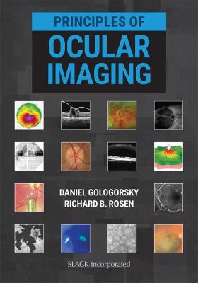
A comprehensive guide for the eye specialist, Principles of Ocular Imaging presents essential information on imaging modalities for ophthalmologists, residents, and fellows.
Ophthalmology and imaging are inextricably intertwined, and practicing eye care professionals need a single definitive source on multiple imaging modalities to reference in clinical practice. Together with their contributors, Drs. Gologorsky and Rosen provide concise but thorough information on the technology and clinical application of 22 imaging modalities unique to ophthalmology, with illustrations and photos throughout that demonstrate how to apply each imaging principle in clinical practice.
Principles of Ocular Imaging is divided into the following subspecialties for easy reference in busy clinical environments:
Oculoplastics: external photography, ptosis visual fields, slit lamp photography, and orbital ultrasonography
Cornea and refractive: corneal topography, confocal microscopy, anterior segment optical coherence tomography (AS-OCT), ultrasound biomicroscopy (UBM), biometry for intraocular lens (IOL) calculations
Glaucoma: visual fields, optical coherence tomography (OCT) in glaucoma
Retina: fundus photography, fluorescein angiography (FA), indocyanine green (ICG) angiography, fundus autofluorescence (FAF), OCT in retina, optical coherence tomography angiography (OCTA), adaptive optics (AO), microperimetry, retinal ultrasonography
Neuro-Ophthalmology: electrophysiology of vision and computed tomography (CT) & magnetic resonance imaging (MRI)
A practical, illustrative guide to ophthalmic imaging, Principles of Ocular Imaging is an indispensable addition to the practicing ophthalmologist's professional library.
Contents:
Dedication
About the Editors
Contributing Authors
Preface
Foreword
Introduction
Section I Oculoplastics
Chapter 1 External Photography
Chapter 2 Ptosis Visual Fields
Chapter 3 Slit Lamp Photography
Chapter 4 Orbital Ultrasonography
Section II Cornea and Refractive
Chapter 5 Corneal Topography
Chapter 6 Confocal Microscopy
Chapter 7 Anterior Segment Optical Coherence Tomography
Chapter 8 Ultrasound Biomicroscopy
Chapter 9 Biometry for Intraocular Lens Calculations
Section III Retina
Chapter 10 Fundus Photography
Chapter 11 Fluorescein Angiography
Chapter 12 Indocyanine Green Angiography
Chapter 13 Fundus Autofluorescence
Chapter 14 Optical Coherence Tomography in Retina
Chapter 15 Optical Coherence Tomography Angiography
Chapter 16 Adaptive Optics
Chapter 17 Microperimetry
Chapter 18 Retinal Ultrasonography
Chapter 19 Electrophysiology of Vision
Section IV Glaucoma
Chapter 20 Visual Fields in Glaucoma
Chapter 21 Optical Coherence Tomography in Glaucoma
Section V Neuro-Ophthalmology
Chapter 22 Computed Tomography and Magnetic Resonance Imaging
Bibliography
Financial Disclosures
Index
PRODUCT DETAILS
Publisher: SLACK Incorporated
Publication date: June, 2021
Pages: 176
Weight: 680g
Availability: Available
Subcategories: Medical Diagnosis, Ophthalmology and Optometry

